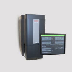
Benchtop SEM
The Benchtop Scanning Electron Microscope (SEM) is an essential tool in biological, medical, and materials science research, offering high-resolution imaging and elemental analysis at the nanoscale. Using electron beam technology, it enables detailed visualization of cell structures, tissue morphology, and biomaterial surfaces, making it valuable in cell biology, pathology, and nanomedicine. In medical research, it helps analyze cell ultrastructure, detect morphological changes due to disease or treatment, and assess biomaterial interactions with tissues. Additionally, in microbiology, it allows the study of bacterial and viral structures, biofilm formation, and host-pathogen interactions.
The Benchtop SEM is also widely used in materials science for characterizing nanoparticles, polymers, and biomedical coatings, as well as in forensic and pharmaceutical research for analyzing drug formulations and contamination. Its ability to perform energy-dispersive X-ray spectroscopy (EDS) enables elemental composition analysis, crucial for studying mineralized tissues, implant materials, and tissue engineering scaffolds. With compact size, ease of use, and rapid imaging capabilities, the Benchtop SEM provides researchers with high-resolution surface analysis, making it an indispensable tool for both biological and material-based investigations.
Core Research Facilities | Site
Ein Karem
Bio Medicine Research Core Facility
Contacts
- Dr. Idit Hazan
- idith@ekmd.huji.ac.il